| |
The Brain: The Body's Control Center
Breathing, blood pressure, heartbeat and the ability to move and feel are controlled by the brain. It's the brain that makes you able to think, to show emotions, and to make judgments. The brain is protected by the skull, tissue, and fluid. 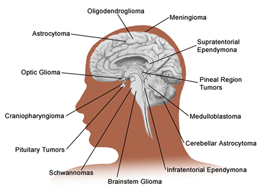
Roles & Functions of the Brain
When you have a tumor, the part of the brain surrounding it may be damaged and the brain can't do its job properly. The brain's right side controls the left side of the body. The left side of the brain controls the body's right side. Some skills and traits occur in more than one section.
Inside the Skull and Protecting the Brain
Under the scalp and the skull, a tough membrane, called the dura, surrounds the brain. Beneath the dura, cerebrospinal fluid (CSF) cushions the brain. Arteries carry nutrients and oxygen-rich blood throughout the brain. Without this blood, brain tissue quickly dies.
Click on the following for information on these specific conditions:
Brain Aneurysm
What is a Brain Aneurysm?
A brain aneurysm is a balloon-like bulge in the wall of a brain artery. If this bulge tears and bleeds, nearby cells may be damaged. A brain aneurysm can occur in an artery wall that is weak or has a defect. An aneurysm is often associated with hardening of the arteries, high blood pressure, heredity, or a head injury.
Symptoms
In most cases, a brain aneurysm has no symptoms until it bleeds or tears. Symptoms include:
- Severe headache – "Worst headache of your life"
- Nausea and vomiting
- Neck stiffness
- Brief blackout
- Confusion or sluggishness
- Vision or speech problems
- Paralysis/weakness on one side
- Clumsiness or jerking movements
Types of Brain Aneurysm
Four main types of Brain Aneurysms exist. Most aneurysms occur where an artery branches, often at the base of the brain. Treatment options vary depending on the type of aneurysm, size, and location.
When an Aneurysm Bleeds
In most cases, the bleeding stops quickly, but if blood that has leaked touches brain cells, the cells may be damaged. Blood in the cerebrospinal fluid (CSF) increases pressure on the brain. Leaked blood may also touch nearby arteries and may cause these arteries to narrow.
Damage to Brain Cells
Blood from an aneurysm can leak into the CSF in the space around the brain (the subarachnoid space). The pool of blood forms a clot, called a hematoma. Blood can irritate, damage, or destroy nearby brain cells and may cause problems with body functions or mental skills. Leaked blood may be removed during surgery.
Fluid Buildup in the Brain
Blood from a torn aneurysm can block CSF circulation, leading to fluid buildup and increased pressure on the brain. The open spaces in the brain (ventricles) then enlarge (called hydrocephalus). It can make a patient lethargic, confused, or incontinent. Fluid may also build up in the brain after surgery. To stop fluid buildup, a drain (called an EVD or extra ventricular drain) may be placed in the ventricles to remove leaked blood and trapped CSF.
Narrowing of Nearby Arteries
An artery may narrow if leaked blood touches it. This response, called vasospasm, may happen up to three to 21 days after an aneurysm bleeds. Vasospasm can decrease blood needed in other parts of the brain and can be fatal. The patient's blood pressure and fluid intake are increased which increases the force of the blood and widens the artery.
Diagnosing a Brain Aneurysm & Treatment Options
Often, the first symptom is a sudden severe headache. Most aneurysm patients describe it as the worst headache of their lives. A physical exam and a health history help to pinpoint the problem. If a brain aneurysm is suspected, special tests can confirm it. Test results help plan treatment, and can include a CT Scan, Spinal Tap and an Arteriogram.
The neurosurgeon will talk with you and may refer to the Hunt-Hess scale, which helps the surgeon assess a patient's condition. Test results and the grade of aneurysm can affect treatment options.
Treatment begins as soon as possible, often within 72 hours of the diagnosis. Either open surgery or an endovascular procedure may be best, but may not reverse any damage already done. The goal is to prevent further bleeding.
 Open Surgery: Clipping, Occlusion and Bypass Open Surgery: Clipping, Occlusion and Bypass
Anesthesia is used during the surgery. The surgeon reaches the brain through the skull. After a scalp incision, small holes are made in the skull. The bone between the holes is cut and lifted away. The dura is peeled back.
Trapped and bloodied CSF may be removed. The surgeon closes off (clips) the aneurysm, or the artery leading to the aneurysm is sealed off (occluded). The dura and the piece of skull are put back in place. A device may be left in one of the small holes which measures pressure inside the skull.
The surgeon may put a clip on the aneurysm where it bulges from the artery. This keeps blood from entering the aneurysm. As a result, future bleeding is prevented and nearby brain tissue is protected from further damage. The surgeon makes sure that the clip is secure before finishing the surgery.
This may be done in surgery with a bypass rather than open surgery. This can be done by the interventional neuroradiologist. A bypass reroutes blood around the occlusion and brings the blood to the part of the brain that had been fed by the damaged artery. A small blood vessel is used for the bypass.
Endovascular Procedure 
It may be best to stop blood flow through the artery leading to the aneurysm, which is called occlusion or embolization. An endovascular procedure may be best for some aneurysms. This is done in an x-ray lab by a specially trained doctor, an Interventional Neuroradiologist.
Under local or general anesthesia, a catheter is guided through the arteries from the groin to the brain, and platinum coils are released into the aneurysm. The coils cause a blood clot to form in the aneurysm, which seals it off.
Brain Tumor
 What is a Brain Tumor? What is a Brain Tumor?
It is a mass of abnormal cells in the brain and there are many types. They may be primary (starting in the brain) or metastatic (traveling to the brain from another site in the body). All brain tumors are either benign or malignant.
What Causes Symptoms?
Along with its location, the way a tumor grows can affect the symptoms you have. A tumor may affect the brain in one or more ways including:
- Destroy normal brain tissue
- Compress normal brain tissue
- Increase pressure in the brain
Symptoms you may have include:
- Headaches that may be worse in the morning
- Trouble thinking, remembering, and/or talking
- Changes in personality
- Vision problems
- Seizures or convulsions causing numbness, weakness, or loss of consciousness
- Paralysis or weakness in one part or on one side of the body
- Loss of balance or lack of coordination
- Nausea and vomiting that may be worse in the morning
Your Medical Evaluation & Treatment Plan
Your doctor will test how well your nervous system is working including checking:
- Thinking/memory skills
- Vision, hearing, talking, and swallowing
- Muscle strength, gait, coordination, and reflexes
- Ability to feel and sense of touch
- Other imaging tests may be necessary
Treatment may include surgery, radiation therapy, Gamma Knife, chemotherapy, or other medications. Treatment may be less involved for certain benign tumors, such as a pituitary tumor. Sometimes a tumor doesn't require treatment yet, but needs to be watched. In this case, it's important to follow-up with your doctor.
If You Have a Biopsy
There are two types of biopsy: open and stereotactic. An open biopsy is done during a craniotomy. With sterotactic biopsy, a sample of the tumor is taken through a small hole made in the skull bone.
Stereotactic Biopsy is often used if a tumor is in a part of the brain that is hard to reach. During this surgical procedure, a special frame may be used to hold the head in place. The biopsy is guided by CT or MRI scans.
Before the biopsy, part of your head may be shaved. You will have anesthesia to numb the part of your head where the surgeon will work, and most patients remain awake.
The surgeon will pass a narrow, hollow needle through the skull bone into the tumor. Cells taken from the tumor will then be sent to a lab to be examined. Risks and complications include bleeding, seizures, and infection.
If You Have a Craniotomy
A craniotomy allows an open biopsy or resection of the tumor to be done and as much of the tumor as possible to be removed.
If You are Given Radiation or Chemotherapy
The goal of radiation therapy is to slow or help control tumor growth. It uses painless x-rays to destroy tumor cells and can with other treatment. If your doctor has selected radiation therapy for you, you may have traditional radiation or sterotactic radiosurgery.
Chemotherapy is a way of treating disease with medications. It may be given as a single medication or a combination. It may be used alone or with surgery or radiation therapy.
Both normal and cancer cells grow and divide, but cancer cells spread out of control. Chemotherapy kills growing cells by interrupting their life cycle. Because it acts on normal cells as well, side effects result. Fast-growing cells – such as those in the hair, digestive system, and blood – are most affected.
It is given in cycles, which allows the body to rest and build healthy cells between treatments. You can receive your therapy in different ways:
- By IV
- By mouth, as a pill
- As a wafer implanted in the brain
Side Effects
Chemotherapy can cause side effects in different parts of your body. Some common short-term side effects and solutions are:
- Nausea or Vomiting
- Low Blood Cell Counts
- Hair Loss
- Mouth Sores
- Long-term side effects and risks are infertility, organ damage, and numbness and tingling in the hands and feet
Carotid Artery Problem
 What is a Carotid Artery Problem? What is a Carotid Artery Problem?
The two common carotid arteries are blood vessels in the neck that supply oxygen-rich blood to your brain. Each carotid artery branches into an internal and external artery. When one of these vessels becomes narrowed, your brain can't get enough oxygen and can lead to a stroke (sometimes called a brain attack).
Symptoms of a Stoke or "Mini-Stroke"
If blood flow to part of your brain stops, even briefly, you may have symptoms of a stroke or "mini-stroke." Seek medical help right away, even if the symptoms last for only a moment. Symptoms include:
- Numbness or weakness in your arms or legs
- Sudden changes in vision or loss of vision in one eye
- Slurring your words
- A facial droop
Open Carotid Arteries
The inside of the artery is open, has no signs of narrowing, is smooth and healthy. Blood flows from your heart to your brain without any problems. Your brain gets all the blood and oxygen it needs.
Narrowed Carotid Arteries
High blood pressure, high cholesterol, diabetes, and other health problems can cause a fatty substance called plaque to build up on the inside of the artery walls. Lifestyle choices such as smoking and a fatty diet can also cause plaque to build up.
The path through the artery is narrowed by plaque buildup. Plaque buildup makes the wall of the artery rough and can cause blood clots to form. Narrowed arteries can prevent some parts of your brain from getting enough blood and oxygen to work normally.
The Dangers of a Narrowed Carotid Artery
Tiny blood clots and bits of plaque can break off and travel through the carotid artery. These are called emboli and can enter the smaller vessels in your brain. If the emboli are large enough, they can block blood flow and cause a stroke.
If You Have a Mini-Stroke or Stroke
Smaller emboli can briefly interrupt blood flow in parts of the brain. This causes a "mini-stroke," also called a transient ischemic attack (TIA). It can last from a few moments to a full day. TIAs are very serious and can be a warning sign of a stroke.
Larger emboli can cut off blood flow to parts of the brain and cause a stroke. Without oxygen-rich blood, that part of the brain dies. Symptoms after a stroke depend on which part of the brain was affected. After a stroke, some people have trouble walking, can't speak, and may die.
When Surgery is Required
If you have mild narrowing, but have had TIAs, you may need surgery. Even if you haven't had any symptoms, your risk may be high if one of your arteries is severely narrowed.
Your Treatment Plan 
If surgery is needed, you'll have a carotid endarterectomy to remove plaque, reopen, and smooth the artery, reducing the chance of emboli forming. Even if you don't need surgery, your doctor may suggest lifestyle changes. Controlling blood pressure, quitting smoking, eating healthier and exercising regularly help reduce your risk. You also may be given medication to help improve your blood flow.
Surgery
A skin incision is made near one of the carotid arteries in your neck. The location and angle aren't always the same. Next, an incision is made in the artery itself.
Your blood may be rerouted for a short time with a shunt. The shunt allows blood to flow to your brain while your doctor works on your artery. If blood flow is strong in your other carotid artery, you may not need a shunt.
The doctor carefully loosens plaque from the artery wall. The plaque is then removed. With the plaque gone, the chance of emboli forming is greatly reduced.
If you have a shunt, it is removed. Your doctor then closes the artery with sutures (stitches). Next, the skin incision is closed and a small tube may be placed in the incision to help with any drainage that may occur. A small bandage will cover the incision.
Craniotomy
 What is a Craniotomy? What is a Craniotomy?
A craniotomy makes a window in the bone covering the brain. This allows a surgeon to reduce pressure beneath the skull. A craniotomy also may be done to remove or repair abnormal structures in the brain.
Why Is a Craniotomy Needed?
Certain problems keep the brain from working correctly. Access to the brain is needed to correct these problems. The problems discussed next are the most common reasons for performing a craniotomy.
Brain Injury - This can result from a direct blow to the head or even whiplash. It can cause tearing, bleeding, and swelling of the brain. The treatment goal is to stop any bleeding and reduce pressure. Blood and damaged tissue may be removed.
Brain Tumor - A tumor is a mass of abnormal cells. The goal is to remove as much of the tumor as possible. Depending on the tumor, other treatments may also be needed. (See page 17 for more details).
Aneurysm - A balloon-like defect in an artery wall. The treatment goal is to control damage and prevent further bleeding. (See page 10 for details).
Arteriovenous Malformation (AVM) - An abnormal tangle of blood vessels which prevents normal blood flow through part of the brain and increases the risk of bleeding into the brain tissue. The treatment goal is to stop blood flow within the AVM and channel it along the normal route.
Diagnosing Your Condition
Your doctor performs certain exams and tests to find the cause of your condition. The results also help learn the precise location and extent of your problem.
Your Medical Exam
Your doctor finds out how well your nervous system is working by checking your:
- Ability to see, hear, walk, and swallow
- Thinking and memory skills
- Muscle strength, coordination, reflexes, and gait
- Ability to feel and sense of touch
- Other imaging tests may be necessary
Reaching the Brain
The surgeon makes an incision in your scalp. Dime-sized burr holes are drilled in the skull and the bone between the holes is cut and lifted away. Then, the surgeon opens the dura exposing the brain. The next step depends on your specific problem.
In some cases, certain nerves may be stimulated while the response in the brain is monitored. This is to make sure that normal brain tissue is not disturbed.
Correcting Your Problem
Brain Injury - The source of bleeding is controlled and blood is removed. Damaged tissue may also be cleaned away.
Brain Tumor - As much of the brain tumor as possible is removed.
Aneurysm - The artery is clipped or sealed at the leak. This prevents more blood from flowing into the brain.
AVM - The abnormal arteries and veins are clipped. This redirects blood flow to normal vessels and prevents the AVM from leaking blood.
Finishing the Craniotomy
When the goal of surgery is met, the dura is closed. In almost all cases, the skull bone is put back. It may be held in place with wire mesh or screw plates. Sometimes blood or fluid remaining in the brain tissue needs to be removed so a drain may be placed through a burr hole for a few days. Most of the time, however, all the burr holes are filled or covered right after surgery. Then the skin incision is closed with stitches or staples.
Other Types of Brain Procedures
The procedures below may be done alone, or they may be performed in addition to a craniotomy. To provide access for shunts or stereotactic surgery, burr holes are made in the skull. Your hospital experience before and after these procedures may be about the same as for a craniotomy.
Head Trauma
What is head trauma?
Head trauma, also known as a traumatic brain injury (TBI), is caused by a blow or jolt to the head that disrupts brain function. Not all blows or jolts to the head result in head trauma. The severity may range from mild, which is a brief change in mental status or consciousness, to severe, an extended period of unconsciousness or amnesia after the injury.
Symptoms
Head trauma can cause a wide range of symptoms. This includes a wide range of functional changes affecting:
- Thinking (memory and reasoning)
- Sensation (touch, taste and smell)
- Language (communication, expression, and understanding)
- Emotions (depression, anxiety, aggression, and social inappropriateness)
Head trauma can also result in epilepsy and increase the risk for conditions such as Alzheimer's disease, Parkinson's disease, and other brain disorders that become prevalent with age.
How is it diagnosed?
The doctor will take into consideration how the injury occurred, as well as the symptoms. A careful neurological evaluation, checking the level of consciousness, reflexes, the size of the pupils and reaction to light, ears, pulse, blood pressure, and breathing rate is performed. Other imaging tests may be necessary.
Surgery
Many types of head trauma may require surgery in order to minimize damage to the brain. These types include primary and secondary head trauma.
Types of Head Trauma
Primary types of brain trauma include direct trauma and indirect trauma. Secondary types of brain trauma include edema, hematoma, hydrocephalus and hygroma. Direct trauma includes any force that penetrates or fractures the skull that may cause severe brain injury such as destructive shock waves, that are sent through the brain matter. Displaced fractures of the skull can also push bone into the brain, causing tissue damage.
Indirect trauma does not involve a direct blow to the head. It includes Shaken Baby Syndrome or severe whiplash. Severe shaking greatly stretches and damages delicate nerve cells, at times causing very significant injury or even death.
 Edema, Hematomas, Hydrocephalus and Hygroma Edema, Hematomas, Hydrocephalus and Hygroma
Edema occurs when the brain swells. This becomes dangerous when the swelling causes a rise in intracranial pressure (ICP) which prevents blood from entering the skull to deliver glucose and oxygen to the brain. If the ICP remains too high for too long, it can be relieved through medication, or in more severe cases, by placing a hole in the skull to drain off some of the high-pressure fluid.
A hematoma is a collection of blood due to tissue injury or the tearing of a blood vessel. CT scans are particularly effective in detecting brain bleeds. Bleeding into the brain after trauma can occur days after the patient is released from the ER or hospital.
A blood clot that develops between the dura and the skull is known as an epidural hematoma. A blood clot that forms between the dura and the brain is known as a subdural hematoma.
Gently resting against the brain itself is a thin, delicate membrane called the arachnoid. Blood leaking into the CSF is known as a sub-arachnoid hemorrhage.
Hydrocephalus and Hygroma are collections of fluid around the brain. If blood gets into the CSF and blocks the spinal fluid absorption sites, spinal fluid will back up into the ventricles, enlarging them, known as hydrocephalus.
If the pressure inside the ventricles becomes excessive, a tube may be inserted into the ventricles to relieve the pressure. A hygroma is a localized fluid buildup usually in the subdural space. If the pressure in the hygroma presses against the brain, surgery may be necessary.
Common Causes of Head Trauma
Common causes of head trauma include traffic accidents, falls, physical assault, and accidents at home, work, outdoors, or while playing sports.
Concussions, Whiplash, and Contusion
A concussion is an alteration of conscious awareness after head trauma. The collection of symptoms following a concussion is called the postconcussion syndrome and include:
- Dizziness
- Nausea
- Vomiting
- Headache
- Disorientation
- Forgetfulness
- Irritability
- Depression
- Mood swings
- Insomnia
- Loss of libido
Most cases of postconcussion syndrome resolve after a few months, but some cases can involve longer term problems.
 Whiplash is the common name for neck sprains due to the neck being thrown forwards and/or backwards at rapid speed. This may cause the fibers of the neck muscles to tear, resulting in pain and a decreased range of movement. Whiplash is the common name for neck sprains due to the neck being thrown forwards and/or backwards at rapid speed. This may cause the fibers of the neck muscles to tear, resulting in pain and a decreased range of movement.
The brain is vulnerable in two ways. The cerebral cortex can become bruised when the head strikes a hard object. Or, the deep white matter can suffer diffuse axonal injury when the head is whiplashed without hitting a hard object. Whiplash may also cause damage to the soft tissue of the spine, such as the tearing of a disc.
Also called a bruise of the brain tissue, a cerebral contusion may be caused by multiple small blood vessels leaking blood into nearby tissue. They commonly heal without medical help.
Seizures & Head Injury
A seizure occurs when nerve cells in the brain rapidly fire electrical discharges at one another. In "grand mal" seizures, this occurs throughout the brain, and results in the patient losing consciousness, falling down, then convulsing.
Seizures may develop immediately after a brain injury or may develop in a delayed fashion, showing up months or even years after the initial trauma. The risk of post traumatic seizures is related to the severity of the injury.
Before Surgery
If there is time before surgery, you may be told to do the following to help your surgery go more smoothly:
- Tell your doctor about any medications you're taking (including aspirin), and ask if you should stop taking them.
- Don't eat or drink anything after midnight before surgery including water, chewing gum, and breath mints.
- If your doctor tells you to take medication the morning of surgery, swallow it with only small sips of water.
- Weeks ahead of time, you may be asked to donate blood for your surgery.
- If you smoke, you should stop now.
- Shortly before the day of your procedure, you will have an exam. This ensures that you are healthy enough for surgery.
|
|